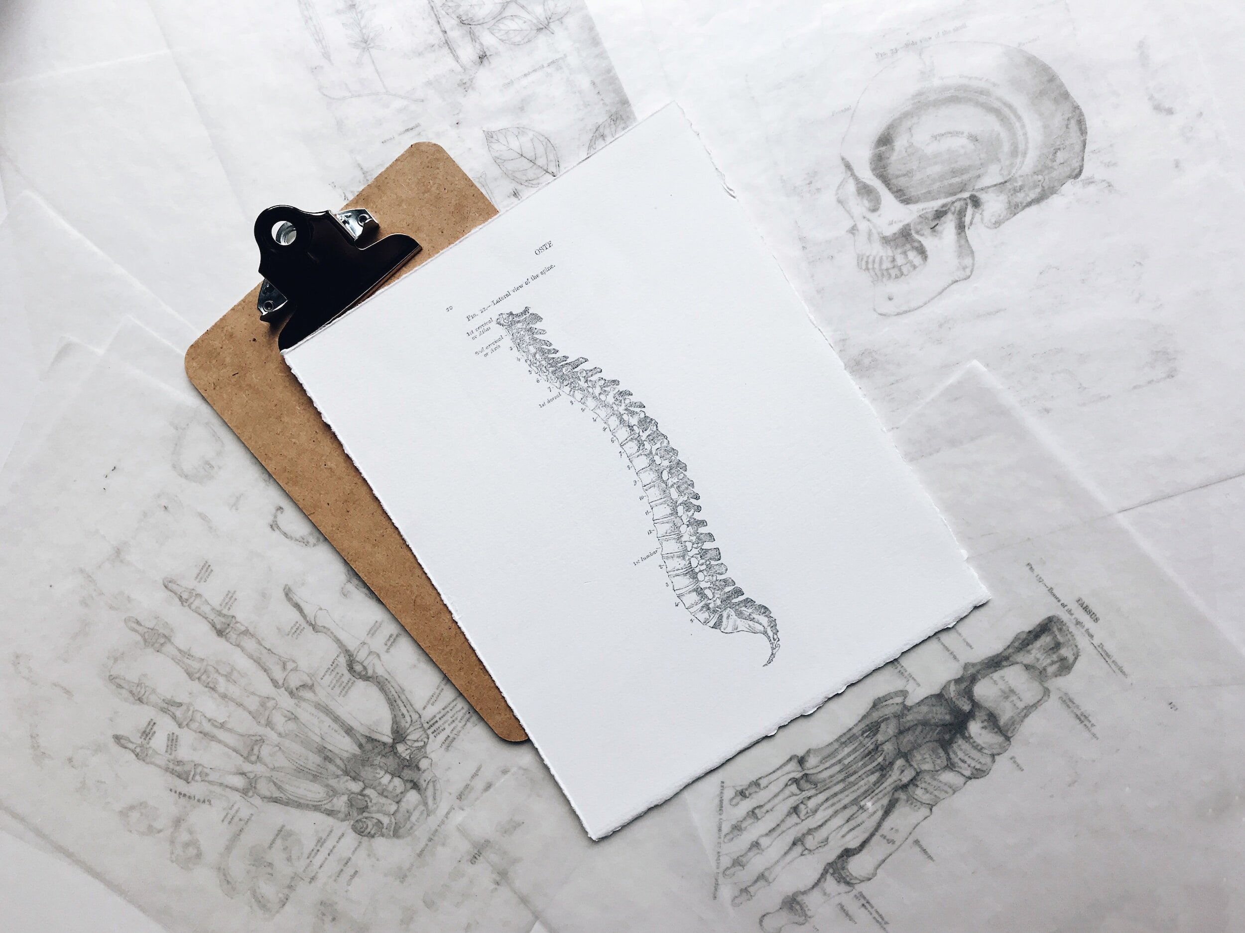
When it comes to our bodies and back pain, we can really get caught up in our diagnosis. “Oh, well I have a bulging disc, so I can’t do X,” or “They showed this level of deterioration at my L4, so I can’t do Y.”
It happens all the time, and I want to emphasize that imaging does not tell the whole story.
I had a client come in the other day, frustrated, holding an image of her X-Ray from a Chiropractor, who’d told her she’d never lift again because of a bulging disc seen on the imaging.
We’d been focusing on treating her right sided back pain, and she handed me the images and the reports from the chiropractor. There were big scary words like maximal deterioration, malaligned, degradation, and the dreaded ‘bulging disc’ all talking about her diagnosis, the compression of the nerves.
I looked at the report, at the image, and my very frustrated patient who looked like the embodiment of Anger from Inside Out.
All of the imaging, and their report, identified the deterioration on the left. What I really wanted to do was chuck that imaging right out the window (too bad my treatment room doesn’t have a window, hah!) This is actually really common; people will find changes on one side, but have symptoms at entirely different levels, or on the other side. In a 10 year longitudinal study, they found no correlation between individuals with age related changes in structure (like disc herniation, disc bulging, or sponylolisthesis) and those individuals with back pain. In otherwords, imaging wasn’t a predictive factor.
Imaging can detect structures. However, there’s not great correlation between who has changes in structures and who has symptoms. Over 50% of individuals over 50 will have “bulging discs” on imaging, but not all of them have back pain! And in another study from this year (Mmm, I love the smell of fresh research in the morning) that looked specifically at patient demographics (like symptoms, age, strength, ect), surgical outcomes, and MRI reports, they found that using MRI imaging in the early stages didn’t have prognostic effects. IE, imaging prior to surgery, including what could be seen on it (potential degenerative changes) wasn’t predictive of how well someone would recover.
Because imaging doesn’t tell the whole story. It’s not going to determine or define your outcomes. This study emphasizes there are so many other things that determine recovery, like age, perceived pain, strength, and sensitivity. Those are strong determining factors; function is predictive, imaging isn’t.
So I talked about it with my client; yes, that’s her imaging. And it by no means guarantees she’ll have back pain, and it certainly doesn’t mean she has to quit crossfit. It’s like looking at a house, seeing a crack in the foundation, and assuming the whole thing was doomed to fail! Haven’t you ever heard of reno work? In 4 weeks, we’d already made major changes. People will use imaging as a diagnostic tool, and as a scare tactic, but it doesn’t capture the whole story, and it doesn’t treat the person in front of you.
Just because you have a disc bulge, some stenosis of your back, a spondylolisthesis, or arthritis doesn’t guarantee you’re going to be symptomatic or have to live with pain. Your strength, your diet, your sleep, and your mindset are all huge factors that tell a much better story than one black and white photo. My client walked out of the room, optimistic, feeling good, empowered in her body, and confident in her understanding of her body. So if someone tries to define you by your imaging when it comes to joints, discs, or muscles, take it with a grain of salt.


